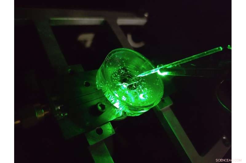
Un prototipo de un microscopio de imágenes de voltaje de diamante construido por físicos de la Universidad de Melbourne. Un pequeño electrodo está suspendido sobre el chip de diamante para probar el rendimiento del dispositivo. Un láser verde que brilla desde abajo proporciona excitación de fluorescencia al chip. Crédito:proporcionado por el autor, Universidad de Melbourne
Podría decirse que el cerebro es una de las estructuras más complejas del universo conocido.
Los continuos avances en nuestra comprensión del cerebro y nuestra capacidad para tratar eficazmente una gran cantidad de enfermedades neurológicas se basan en sondear los microcircuitos neuronales del cerebro con cada vez mayor detalle.
Una clase de métodos para estudiar los circuitos neuronales se denomina formación de imágenes de voltaje. Estas técnicas nos permiten ver el voltaje generado por las neuronas de nuestro cerebro, lo que nos dice cómo se desarrollan, funcionan y cambian las redes de neuronas con el tiempo.
Hoy en día, las imágenes de voltaje de las neuronas cultivadas se realizan utilizando conjuntos densos de electrodos en los que se cultivan las células, o mediante la aplicación de tintes emisores de luz que responden ópticamente a los cambios de voltaje en la superficie de la célula.
Pero el nivel de detalle que podemos ver usando estas técnicas está restringido.
Los electrodos más pequeños no pueden distinguir de forma fiable las neuronas individuales, unas 20 millonésimas de metro de ancho, por no hablar de la densa red de conexiones a nanoescala que se forma entre ellas, y no se han realizado avances tecnológicos significativos en esta área durante más de dos décadas.
Además, cada electrodo requiere su propia conexión por cable y amplificador, lo que impone limitaciones significativas en la cantidad de electrodos que se pueden medir simultáneamente.
Los tintes pueden superar estas limitaciones al generar imágenes del voltaje de forma inalámbrica como luz, lo que significa que la electrónica compleja puede ubicarse lejos de las celdas dentro de una cámara.
El resultado es una alta resolución sobre grandes áreas, capaz de distinguir cada neurona individual en una gran red. Pero aquí también hay limitaciones, las respuestas de voltaje de los tintes de última generación son lentas e inestables.
Nuestra investigación reciente publicada en Nature Photonics , explora un nuevo tipo de plataforma de imágenes de voltaje escalable, de alta resolución y de alta velocidad creada con el objetivo de superar estas limitaciones:un microscopio de imágenes de voltaje de diamante.
Desarrollado por un equipo de físicos de la Universidad de Melbourne y la Universidad RMIT, el dispositivo utiliza un sensor de diamante que convierte las señales de voltaje en su superficie directamente en señales ópticas, lo que significa que podemos ver la actividad eléctrica en el momento en que sucede.
The conversion uses the properties of an atom-scale defect in the diamond's crystal structure known as the nitrogen-vacancy (NV).
NV defects can be engineered by bombarding the diamond with a nitrogen ion beam using a special type of particle accelerator. The fabrication of the sensor begins with using this process to create a high-density, ultra-thin layer of NV defects close to the diamond's surface.
You can think of each NV defect as a bucket that holds up to two electrons. When this bucket is empty, the NV defect is dark. With one electron, the NV defect emits orange light when illuminated by a laser—this property is known as fluorescence. With two electrons, the color of the fluorescence becomes red.
A previously discovered property of NV defects is that the number of electrons they hold—and the resulting fluorescence—can be controlled with a voltage. Unlike dyes, the voltage response of an NV defect is very fast and stable.
Our research aims to overcome the challenge of making this effect sensitive enough to image neuronal activity.
On the diamond's surface, the crystal structure ends with a layer one atom thick, made up of hydrogen and oxygen atoms. The NV defects closest to the surface are the most sensitive to changes in voltage outside the diamond, but they are also highly sensitive to the atomic makeup of the surface layer.
Too much hydrogen and the NVs are so dark that the optical signals we are looking for cannot be seen. Too little hydrogen and the NVs are so bright that the small signals we are after are completely washed out.
So, there's a "Goldilocks' zone" for voltage imaging, where the surface has just the right amount of hydrogen.
To reach this zone, our team developed an electrochemical method for removing hydrogen in a controlled way. By doing this, we've managed to achieve voltage sensitivities two orders of magnitude better than what has been previously reported.
We tested our sensor in salty water using a microscopic wire 10-times thinner than a human hair. By applying a current, the wire can produce a small cloud of charge in the water above the diamond. The formation and subsequent diffusion of this charge cloud produces small voltages at the diamond surface.
By capturing these voltages through a high-speed recording of the NV fluorescence, we can determine the speed, sensitivity and resolution of our diamond imaging chip.
We were able to further boost sensitivity by patterning the diamond's surface into 'nanopillars'—conical structures with the NV centers embedded in their tips. These pillars funnel the light emitted by the NVs towards the camera, dramatically increasing the amount of signal we can collect.
With the development of the diamond voltage imaging microscope for detecting neuronal activity, the next step is the recording of activity from cultured neurons in vitro—these are experiments on cells grown outside their normal biological context, otherwise known as test-tube or petri-dish experiments.
What differentiates this technology from existing state-of-the-art in vitro techniques is the combination of high spatial resolution (on the order of a millionth of a meter or less), large spatial scale (a few millimeters in each direction—which for a network of neurons in mammals is quite vast), and complete stability over time.
No other existing system can simultaneously offer these three qualities, and it's this combination that will allow our made-in-Melbourne technology to make a valuable contribution to the work of neuroscientists and neuropharmacologists globally.
Our system will aid these researchers in pursuing both fundamental knowledge and the next generation of treatments for neurological and neurodegenerative diseases. New method enables long-lasting imaging of rapid brain activity in individual cells deep in the cortex